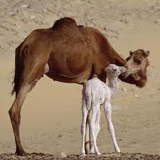
Age estimation of camel in Nigeria using rostral dentition
Keywords:
Age, Camel, Dentition, Estimation, NigeriaAbstract
The study aimed at providing information in estimating the age of camel using rostral dentition through the phenomenon of teeth eruption and wearing, thought, only way to age an animal accurately is to know the date of birth but where these records are not available various anatomical features are used to estimate age. A total of 1100 camels of both sex were used for the purpose of the study. Records were obtained between April to July 2010 on daily visit to the Sokoto metropolitan abattoir. Investigation showed that at birth, there were no teeth, at 9 month, all the deciduate teeth have erupted. At 4 years, all the deciduate incisors and canine have worn down. At 7 years, all the permanent incisors and canine teeth have erupted. At 12 years, all the permanent incisors are in wear, while At 15 years, all the permanent incisors and canine teeth have worn down. At 20 years, all the permanent teeth are down and clearly separated from each other.
References
Ainamo, J., 1970. Morphogenetic and Functional Characteristics of Coronal Cementum in Bovine teeth,
Scandinavian Journal of Dental Research, 78,378-386.
de-Lahunta, A., 1986. Applied Veterinary Anatomy, 5th edition, W.B. Sanders Company, Pp. 5-15.
Arnautovic, I., Abdelmagid, A.M., 1974. Anatomy and mechanism of distension of gula of one humped camel. Acta
Anatomica, 88, 115-124.
Berkowitz, B.K.B., Moxham, B., 1981. Development of dentition: Early stages of tooth development. In: Dental
Anatomy and Embryology, ed. JW Osborn, Blackwell Scientific Publications, Oxford, pp. 166-174.
Ferguson, M., 1990. The dentition throughout life. In: The Dentition and Dental Care, Vol. 3, ed. RJ Elderton, Oxford
Heinemann Medical Books, Oxford, pp. 1-18.
Food and Agricultural Organization, (FAO)., 1990. Animal Health Year Book, Rome: FDA; P. 3.
Harvey, C.E. 1985. Function and Formation of oral cavity. In Harvey C.E. edited by Veterinary Dentistry
Philadelphia, P.A: W.B. Sanders Company, Pp. 5-22.
Jamalar, M.A., 1996. Comparative Anatomy of the bony 8 years of the camel (camelus dromedarus). Indian
Veterinary Journal, 37,225-239.
Latshaw, W.K., 1987. Face, mouth and pharynx. In: Veterinary Developmental Anatomy – A clinically Oriented
Approach, ed. WK Latshaw, BC Decker, Toronto
Malie, M., Smuts, S., Bezuidenhout, 1987. Anatomy of the dromedarius camel. Clarenden press, Oxford.
Saghiri, A., Driencourt, M.A., 1999. Seasonal effects on fertility and ovarian follicular growth and maturation in
camels (Camelus dromedarius). Journal of Animal Science, 55, 223-237.
Swift, J.J., 1979. The development of livestock trading in a nomad pastoral economy: The Somalia case. In Pastoral
Production and Society, Cambridge University Press, Cambridge, UK. pp 447-465.
Watson, R.M., 1969 .A census of the domestic stock of Inort Eastern Province, Kenya (Mimeo), Nairobi. p 135.
Wilson, R.T., 1984. Research in camel. A look at the past. Some pointers for the future in Animal Production in the
tropics. Yousek, 1st edition, New York, USA.
Yasin, S.A., Walhid, A., 1975. Pakistan Camels – A Preliminary Survey. Agric. Pakistan, Chapter 8, Pp. 289-297.
Zeidan, A.E.B., 1999. Effect of age on some reproductive traits of the male one-humped camels (Camelus
dromedarius). Zagazig Veterinary Journal, 27, 126-133.

Downloads
Published
How to Cite
Issue
Section
License
Copyright (c) 2013 A. Bello, M. L. Sonfada, A. A. Umar, M. A. Umaru, S. A. Shehu, S. A. Hena, J. E. Onu, O. O. Fatima

This work is licensed under a Creative Commons Attribution-NonCommercial-NoDerivatives 4.0 International License.


