Genetic mice models of Parkinson’s disease
Keywords:
Parkinson's disease, Dopaminergic neurons, Neurodegeneration, Substantia nigra pars compacta, Gene, MutationAbstract
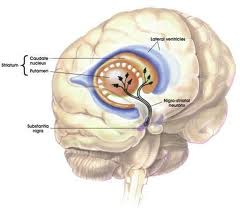
The loss of dopaminergicneurons in the substancia nigra pars compacta leads to the characteristicsymptoms of Parkinson's disease (PD), whose Lewy bodies is a pathologic signalmost always found. Various monogenic forms account for a minority of cases ofPD, but have provided crucial insight into disease mechanism. However,genetically faithful models have not been exposed to putative toxicants in amanner that is clearly relevant to human exposures, and most of studies haveused conventional genetically modified animals and convenient dosing paradigms.Better translation between preclinical, neuropathologic animal model and,clinical research would be important for future clinical trials.References
Andres-Mateos, E., Mejias, R., Sasaki, M., Li, X., Lin, B.M., Biskup, S., 2009. Unexpected lack of hypersensitivity in LRRK2 knock-out mice to MPTP (1-methyl-4-phenyl-1,2,3,6-tetrahydropyridine). J. Neurosci. 29, 15846–50.
Berthet, A., Bezard, E., Porras, G., Fasano, S., Barroso-Chinea, P., Dehay, B., 2012. L-DOPA Impairs Proteasome Activity in Parkinsonism through D1 Dopamine. Receptor J. Neurosc. 32(2), 681– 691.
Chesselet MF, Richter F. Modelling of Parkinson´s disease in mice. Lancet. Neurol. 10, 1108-18.
De Lau, K.M., Breteler, M.M., 2011. Epidemiology of Parkinson´s disease. Lancet Neurol 2006; 5: 525-35.
Dawson, T.M., Ko, H.S., Dawson, V.L., 2010. Genetic animal models of Parkinson´s disease. Neuron. 66, 646-61.
Greggio, E., Zambrano, I., Kaganovich, A., Beilina, A., Taymans, J.M., Daniels, V., 2008. The Parkinson disease-associated Leucine-rich Repeat Kinase 2 (LRRK2) is a dimer that undergoes intramolecular autophosphorylation. J. Biol. Chem. 283, 16906–14.
Hirsh, E., Graybiel, A.M., Agid, Y.A., 1988. Melanized dopaminergic neurons are differentially susceptible to degeneration in Parkinson's disease. Nature. 334, 345-8.
Lewis, S.J.G., Barker, R.A., 2009. Understanding the dopaminergic deficits in Parkinson’s disease: Insights into disease heterogeneity. J. Clini. Neurosci. 16, 620-65.
Qing, X., Sushila, S., Chenjian, L., 2012. Mouse models for LRRK2 Parkinson’s disease. Parkinsonism. Relat. Disord. 18S1, S186-S189.
West, A.B., Moore, D.J., Biskup, S., Bugayenko, A., Smith, W.W., Ross, C.A., 2005. Parkinson’s disease-associated mutations in leucine-rich repeat kinase 2 augment kinase activity. Proc. Natl. Acad. Sci. USA, 102, 16842–7.
Published
How to Cite
Issue
Section
Copyright (c) 2013 R. Bolaños-Jiménez, C. Escamilla-Ocañasb, H. Martínez-Menchacab, G. Rivera-Silva, O. F. Chacon-Camacho, J. D. Carrillo-Ruíz

This work is licensed under a Creative Commons Attribution-NonCommercial-NoDerivatives 4.0 International License.

